TOPIM 2013 - 7th Winter Conference

IMAGING the HALLMARKS of CANCER

“Neither the sun nor death can be looked at with a steady eye"
wrote Francois de La Rochefoucauld (1613-1680).


Today's in vivo images of cancer demonstrate that cancer is no longer synonym of death but still linger somewhere in between blindness and clarity. The next stage in cancer imaging will be to produce and interpret in vivo images of cancer according to the biological hallmarks of cancer defined by Hanahan and Weinberg: sustained proliferative signaling, evasion from growth suppressors and immune destruction, enabling of replicative immortality, tumor-promoting inflammation, activation of invasion and metastasis, induction of angiogenesis, genomic instability and mutation, resistance to cell death and deregulation of cellular energetics.


THANKS to all of you who contributed a great conference!

"Congratulations for a great conference! This conference was excellent not only for the science but also for the friendly athmosphere, the professionel organization, the inspiring discussions [...]"

TOPIM 2013 participant

The 10 biological hallmarks of cancer defined by Hanahan and Weinberg defined also the topics for TOPIM 2013. Per topic some speakers were invited to give a talk within their field of expertise (seniors) and some talks were given by junior scientists – choosen from the submitted abstracts. 

The programme started on Monday morning with a 3h introductoryKeynote lecture by Robert Gillies. Further Keynote lectures were given by Douglas Hanahan and John Condeelis.

Keynote Lectures

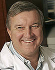
Douglas Hanahan

Douglas Hanahan is Director of the Swiss Institute for Experimental Cancer Research - ISREC in Lausanne. In 2000, Douglas Hanahan and Robert Weinberg published a very influential paper called “The Hallmarks of Cancer” in the journal Cell in which they suggested 6 hallmark traits that confer malignancy on cells. This classic paper was recently updated (Hanahan and Weinberg 2011) to add two additional “emerging” hallmarks and two “enabling characteristics.”

ABSTRACT Douglas Hanahan

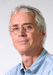
John Condeelis

John Condeelis is Professor & Co-Chair of Anatomy & Structural Biology at the Albert Einstein College of Medicine in New York.  His research interests are in optical physics, cell biology and biophysics, cancer biology and mouse models of cancer. He and his collaborators developed the multiphoton imaging technology and animal models used to identify invasion and intravasation microenvironments in mammary tumors. This led to the discovery of the paracrine interaction between tumor cells and macrophages in vivo, and the role of macrophages in the migration of tumor cells and their dissemination from primary tumors via blood vessels to distant metastatic sites read more

ABSTRACT John Condeelis

Robert Gillies

Robert J. Gillies is vice-chair of Radiology, director of the Department of Imaging Research and co-leader of the Experimental Therapeutics Programme at the Moffitt Cancer Center in Tampa, USA. His research is focused on functional and molecular imaging of cancer: tumour microenvironment, imaging biomarkers for therapy, tumour targeting ligands. He is a member of the Cell and Molecular Imaging Probes - CMIP study section at NIH and a member of IRG-A Cancer Center Grants parent committee at NCI.

ABSTRACT Robert Gillies
Poster Session and Award

Two guided poster sessions were held this year in Les Houches. The best poster presenters had the opportunity to give a talk the following day. The winners:

Sylvia Le Dévédec from Leiden on "Kinome wide screening for regulators of focal adhesions dynamics identifies PFKFB2 as a novel determinant of breast cancer progression" (poster# 5)

Christoph Griessinger from Tuebingen on "Influence of the administration route on the treatment efficiency of tumor-associated antigen (TAA) specific Th1 cells in an animal model for pancreatic cancer" (poster# 24)


TOPIM 2013 was also co-funded by the FP7 project INMiND - Imaging of Neuroinflammation in Neurodegenerative Diseases.